
HFE Gene
In 1976, hereditary hemochromatosis was found to be closely linked to MHC class I molecules (Simon et. al., 1976). This breakthrough led to the discovery of the HFE gene, which was found to encode proteins with structural similarities to other MHC class I molecules (Fleming et. al., 2005). The HFE gene was proven to be the defective gene in the disease when the knockout of the mouse HFE gene resulted in iron overload (Zhou et. al., 1998).
In nearly all patients with MHC-linked hemochromatosis, a single nucleotide change of tyrosine into cysteine at amino acid 282 was identified. A second mutation of an aspartate substitution in place of histidine at amino acid 63 was identified in the HFE gene (Fleming et. al., 2005).
The C282Y mutation is seen in the majority of hemochromatosis patients of northern European ancestry, while the H63D mutation appears in different ethnic populations (Rochette et. al., 1999). Population studies suggest that the C282Y mutation occurred on an ancestral haplotype roughly 2000 years ago (Rochette et. al., 1999). In the patients of northern European ancestry, approximately 80 to 90% of the subjects with hemochromatosis were homozygous for C282Y (Powell et. al., 2000). However, many of the C282Y homozygotes do not develop clinically significant levels of iron overload early on, indicating that there is incomplete entrance of the C282Y mutation (Fleming et. al., 2005). Incomplete penetrance of the C282Y mutation is due to genetic modifiers. Similarly, homozygosity of the H63D mutation does not result in clinically significant levels of iron overload. However, heterozygosity of the H63D mutation with the C282Y mutation is increasingly found in patients with iron overload (Bacon et. al., 2001). The most prevalent gene mutations seem to be homozygous C282Y and heterozygous C282Y/H63D.
HFE Protein
The HFE gene encodes for the HFE protein, which is structurally similar to MHC class I molecules. Although the HFE protein lacks immunological functions, like other MHC class I molecules, it is physically associated with β2-microglobulin (β2M) (Feder et. al., 1997). As seen in Figure 1a, an eight-stranded antiparallel β-sheet with two α-helices on top comprises the α1–α2 superdomain (residues 1-180), which is positioned on top of the α3 domain (residues 181–275) and β2M (Bennett et. al., 2000). The C282Y mutation leads to the disruption of the disulfide bond in the α3 domain necessary for the association with β2M (Bennett et. al., 2000). As a result, the C282Y mutant protein is abnormally processed, causing accelerated degradation and reduced cell surface expression (Parkkila et. al., 2000). The association with β2M is necessary for normal HFE protein function (Santos et. al., 1996).
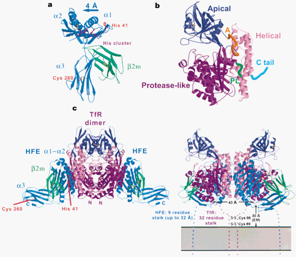
The three domains are presented in Figure 1b. The domains consist of a protease-like domain (residues 121-188 and 384-606), an apical domain (residues 189-383), and a helical domain (residues 607-760) (Bennett et. al., 2000). Figure 1c reveals that the protein complex structure consists of a transferrin receptor 1 (TfR1) homodimer with one HFE molecule bound to each chain, forming a two-fold symmetric complex (Bennett et. al., 2000). The primary interaction occurs between the HFE α1–α2 platform and the two helical domains of TfR1 (Bennett et. al., 2000). The H63D mutation alters the α1 domain into a loop rearrangement to avoid proper residue interactions (Lebrón et. al., 1998).
In order to understand how the HFE protein plays a role in iron overload, the interactions between HFE protein, TfR1, and transferrin (Tf) were characterized (Lebrón et. al., 1998). By binding to TfR1, HFE reduces the affinity of TfR1 binding with iron (Fe). With a mutated HFE protein, as in hemochromatosis, the binding of the HFE-TfR1 complex occurs at a lower affinity (Lebrón et. al., 1998). After these discoveries, it was then determined that HFE also binds transferrin receptor 2 (TfR2), functioning to sense iron levels in the cell (Goswami et. al., 2006). When high levels of Fe are present, HFE is displaced from TfR1 and binds to TfR2, signaling a cascade for hepcidin production (Goswami et. al., 2006).
Hepcidin Protein and Iron Overload
Hepcidin plays a role in iron regulation and is considered to be the master iron regulatory hormone. The involvement of hepcidin in iron homeostasis was first observed in liver hepcidin mRNA expression in mice, which increased with dietary iron loading (Pigeon et. al., 2001). A hepcidin knockout established its critical role as a negative regulator of iron absorption (Pigeon et. al., 2001). Cells that are involved in systemic iron regulation are shown in Figure 2.
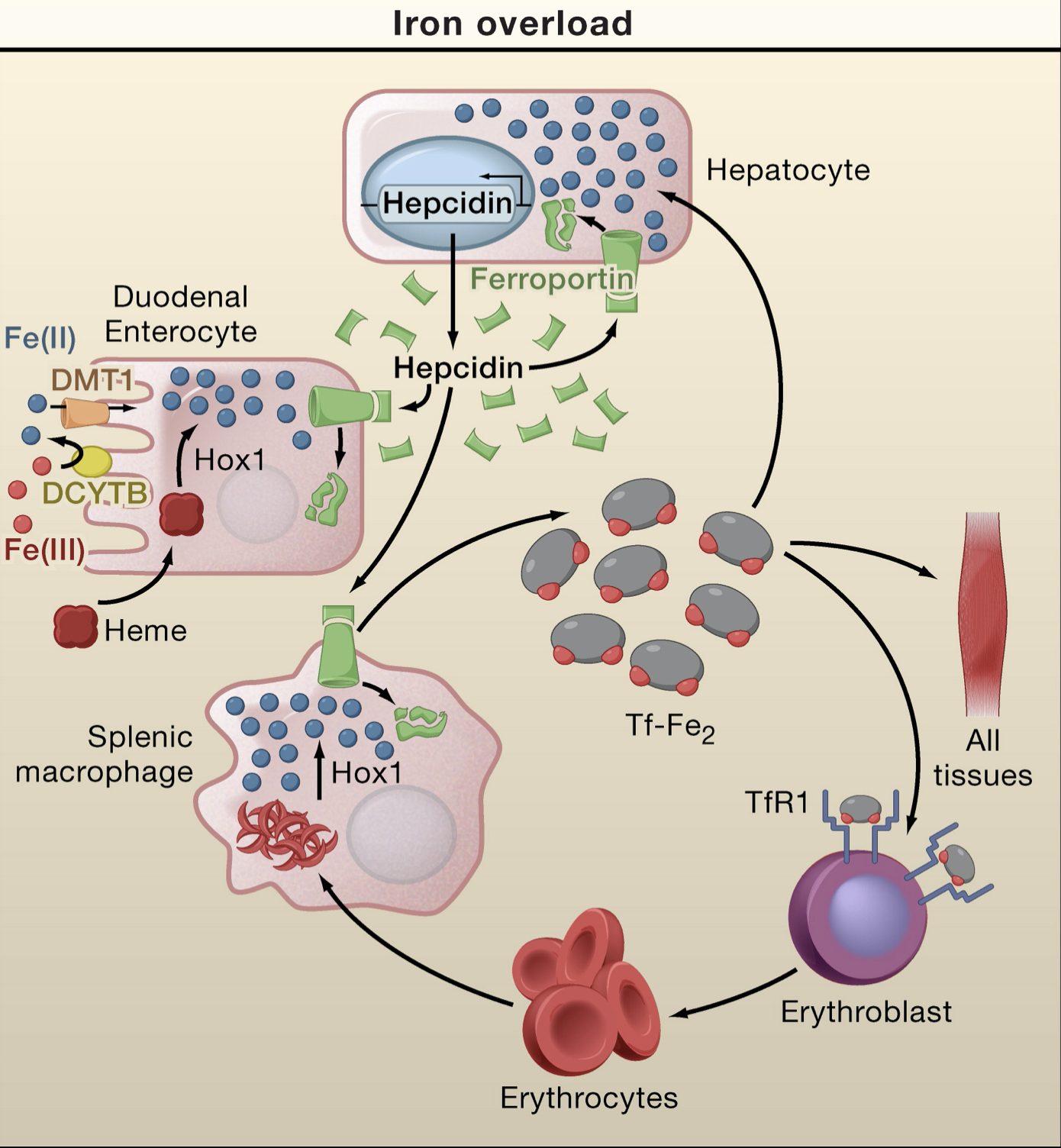
At the apical membrane of enterocytes, DCYTB reduces Fe(III) to Fe(II) causing divalent metal transporter 1 (DMT1) to take up iron from the lumen of the duodenum (Hentze et. al., 2010). At the basolateral membrane, ferroportin works with hephaestin which oxidizes Fe(II) to Fe(III) (Hentze et. al., 2010). Tf-Fe (indicated by red dots) supplies iron to all cells by binding to TfR1 and undergoing endocytosis. Hepatocytes sense this transferrin saturation and release hepcidin. The transferrin saturation is reduced with high levels of hepcidin because the higher concentration of the protein inhibits ferroportin-mediated iron export by triggering internalization and degradation of the complex (Hentze et. al., 2010). The mechanism for hepcidin release is shown in Figure 3.

The HFE protein is displaced from TfR1 in the presence of high concentrations of Tf-Fe in order to promote its interaction with TfR2 (Hentze et. al., 2010). HFE regulates hepcidin expression through the bone morphogenetic protein (BMP) pathway (Muckenthaler et. al., 2014). The HFE-TfR2 complex activates hepcidin transcription via ERK/MAPK and BMP/SMAD signaling. At the plasma membrane, phosphorylation of receptor activated SMAD proteins is induced by the interaction between the BMP coreceptor hemojuvelin (HJV) and type I and type II BMP receptors (BMPR) (Hentze et. al., 2010). This interaction causes the formation of active transcriptional complexes involving the co-SMAD factor SMAD4 (Hentze et. al., 2010)
Further research has elucidated missing steps in the hepcidin expression pathway, shown in Figure 4. HFE binds to ALK3, a type of BMP receptor, to promote cellular expression and inhibit ubiquitination (Wu et. al., 2014). BMP6, HJV, and ALK3 form a complex that induces phosphorylation of the previously mentioned complex, thereby inducing expression of hepcidin (Wu et. al., 2014).

HFE proteins with the C282Y mutation cannot induce the BMP pathway to express hepcidin, since the mutated protein struggles to facilitate ALK3 transport to the cell surface (Wu et. al., 2014). The inability to transport ALK3 causes low levels of hepcidin, which inhibits the ubiquitination and degradation of ferroportin (Wu et. al., 2014). As a result, iron is continuously absorbed by enterocytes and recycled from macrophages, creating the iron deposits that are seen in hemochromatosis.
a
Normal Function of Key Players
Previous: Identification and Characterization
Next: Effects, Detection, and Treatments
a
References
Bacon, B. R. Hemochromatosis: Diagnosis and Management. Gastroenterology 2001, 120 (3), 718–725.
Bennett, M. J.; Lebrón, J. A.; Bjorkman, P. J. Crystal Structure of the Hereditary Haemochromatosis Protein HFE Complexed with Transferrin Receptor. Nature 2000, 403 (6765), 46–53.
Feder, J. N.; Tsuchihashi, Z.; Irrinki, A.; Lee, V. K.; Mapa, F. A.; Morikang, E.; Prass, C. E.; Starnes, S. M.; Wolff, R. K.; Parkkila, S.; Sly, W. S.; Schatzman, R. C. The Hemochromatosis Founder Mutation in HLA-H Disrupts Β2-Microglobulin Interaction and Cell Surface Expression. J. Biol. Chem. 1997, 272 (22), 14025–14028.
Fleming, R. E.; Britton, R. S.; Waheed, A.; Sly, W. S.; Bacon, B. R. Pathophysiology of Hereditary Hemochromatosis. Semin Liver Dis 2005, 25 (4), 411–419.
Goswami, T.; Andrews, N. C. Hereditary Hemochromatosis Protein, HFE, Interaction with Transferrin Receptor 2 Suggests a Molecular Mechanism for Mammalian Iron Sensing. J. Biol. Chem. 2006, 281 (39), 28494–28498.
Hentze, M. W.; Muckenthaler, M. U.; Galy, B.; Camaschella, C. Two to Tango: Regulation of Mammalian Iron Metabolism. Cell 2010, 142 (1), 24–38.
Lebrón, J. A.; Bennett, M. J.; Vaughn, D. E.; Chirino, A. J.; Snow, P. M.; Mintier, G. A.; Feder, J. N.; Bjorkman, P. J. Crystal Structure of the Hemochromatosis Protein HFE and Characterization of Its Interaction with Transferrin Receptor. Cell 1998, 93 (1), 111–123.
Muckenthaler, M. U. How Mutant HFE Causes Hereditary Hemochromatosis. Blood 2014, 124 (8), 1212–1213.
Parkkila, S.; Parkkila, A. K.; Waheed, A.; Britton, R. S.; Zhou, X. Y.; Fleming, R. E.; Tomatsu, S.; Bacon, B. R.; Sly, W. S. Cell Surface Expression of HFE Protein in Epithelial Cells, Macrophages, and Monocytes. Haematologica 2000, 85 (4), 340–345.
Pigeon, C.; Ilyin, G.; Courselaud, B.; Leroyer, P.; Turlin, B.; Brissot, P.; Loréal, O. A New Mouse Liver-Specific Gene, Encoding a Protein Homologous to Human Antimicrobial Peptide Hepcidin, Is Overexpressed during Iron Overload. J. Biol. Chem. 2001, 276 (11), 7811–7819.
Powell, L. W.; Subramaniam, V. N.; Yapp, T. R. Haemochromatosis in the New Millenium. Journal of Hepatology 2000, 32, 48–62.
Rochette, J.; Pointon, J. J.; Fisher, C. A.; Perera, G.; Arambepola, M.; Arichchi, D. S.; De Silva, S.; Vandwalle, J. L.; Monti, J. P.; Old, J. M.; Merryweather-Clarke, A. T.; Weatherall, D. J.; Robson, K. J. Multicentric Origin of Hemochromatosis Gene (HFE) Mutations. Am. J. Hum. Genet. 1999, 64 (4), 1056–1062.
Santos, M.; Schilham, M. W.; Rademakers, L. H.; Marx, J. J.; de Sousa, M.; Clevers, H. Defective Iron Homeostasis in Beta 2-Microglobulin Knockout Mice Recapitulates Hereditary Hemochromatosis in Man. J. Exp. Med. 1996, 184 (5), 1975–1985.
Simon, M.; Bourel, M.; Fauchet, R.; Genetet, B. Association of HLA-A3 and HLA-B14 Antigens with Idiopathic Haemochromatosis. Gut 1976, 17 (5), 332–334.
Wu, X.; Wang, Y.; Wu, Q.; Cheng, W.-H.; Liu, W.; Zhao, Y.; Mayeur, C.; Schmidt, P. J.; Yu, P. B.; Wang, F.; Xia, Y. HFE Interacts with the BMP Type I Receptor ALK3 to Regulate Hepcidin Expression. Blood 2014, 124 (8), 1335–1343.
Zhou, X. Y.; Tomatsu, S.; Fleming, R. E.; Parkkila, S.; Waheed, A.; Jiang, J.; Fei, Y.; Brunt, E. M.; Ruddy, D. A.; Prass, C. E.; Schatzman, R. C.; O’Neill, R.; Britton, R. S.; Bacon, B. R.; Sly, W. S. HFE Gene Knockout Produces Mouse Model of Hereditary Hemochromatosis. PNAS 1998, 95 (5), 2492–2497.

2 Responses
Hey Iffat! Thanks for writing such an interesting and well-organized blog! Hemochromatosis is prevalent in my family, so it’s been especially rewarding to learn about how this condition works. It’s funny that you mention phlebotomy on your treatments page– my loved ones and I donate blood regularly as a way to mediate the iron buildup while helping a good cause! After reading this page on the molecular details involved with hemochromatosis, I wanted to hear your thoughts on the C282Y mutation you mention toward the end of the text. You say that the mutated HFE proteins are unfit for ALK3 transport, which then causes a cascade of problems. Have you come across any papers that discuss why substituting a tyrosine for a cystine is specifically the issue? Is the sulfur in C involved in binding in a way that Y can’t support? Or perhaps is the ring in Y a steric block?
Thanks for your comment, Em! You bring up a great point! With our biochemical background, we have become so wired to think about how these mechanisms are occurring between the specific amino acid structures. The questions you have posed were actually very similar to what I had been thinking of throughout this whole project. I haven’t been able to find any papers that discuss the chemical/structural reasons behind the effects of the mutation. I think that is more due to the fact that the details of how the mutations of hemochromatosis mechanistically affect the overall BMP-SMAD pathway (which the ALK3 is a part of) is unknown.
Typically, HFE associates with ALK3, inhibiting ALK3 ubiquitination and proteasomal degradation, increasing ALK3 protein expression and accumulation on the cell surface. The mutations typically affect ALK3 in a way that results in a decrease of the protein accumulation on the cell surface. The ALK are serine-threonine kinase receptors, which are enzymes that phosphorylate the OH of serine or threonine. Even though tyrosine has an OH in its side chain, I suppose the benzene ring of the tyrosine may pose as an steric issue. You also brought up the sulfur binding of the cysteine, which may also have better polar interactions in the active site over the tyrosine. The details are still unknown, but more current information on the BMP-SMAD pathway can be found here.
The consequences of the mutations are widely known in the scientific community, but the mechanism of what the mutations do is unclear.